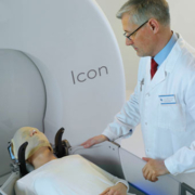
Research into the use of Gamma Knife to treat primary brain tumours is ever evolving. In August 2022 the Journal of Neurosurgery (JNS) published the results of a feasibility study in a clinical article called, ‘Conventionally fully fractionated Gamma Knife Icon re-irradiation of primary recurrent intracranial tumors: the first report indicating feasibility and safety’.

Ian Paddick, Chief Physicist for Amethyst UK comments on the recently published article. He said; “This collection of case studies demonstrates the feasibility and potential safety of using Gamma Knife to deliver conventionally fractionated salvage treatments to recurrent tumours close to organs at risk that have received doses close to, or up to full tolerance.

“In addition, the article goes on to remind us that we have animal and retrospective clinical data that shows that brain tissue can recover significantly after irradiation – perhaps 75% in two years. This allows for reirradiation. Therefore, there may be room to be more aggressive when clinically needed.”

Why use Gamma Knife for this sort of treatment?

Gamma Knife is ideally suited to irradiation of complex targets to low doses. It can, for example, deliver complex treatment plans in 2Gy fractions in around 10 minutes, making it as efficient as a Linac. The feasibility study highlighted in the JNS article used a median number of 28 fractions. Ian Paddick concluded; “Gamma Knife’s excellent conformity and gradient is better suited to target tumours surrounded by previously irradiated tissue.”

Gamma Knife Imaging

Cerebral arteriovenous malformations (AVMs) arise when an abnormal tangle of blood vessels occurs in the brain as a result of an abnormal connection between an artery and vein. These can have serious consequences such as haemorrhaging, stroke, brain damage and seizures. Gamma Knife radiosurgery represents a massive improvement in the treatment options for AVMs, as a targeted and precise, non-invasive approach.

What are the treatment options available for AVMs?

The three treatment options are endovascular embolization (“gluing”), neurosurgical resection (surgery) and stereotactic radiosurgery (Gamma Knife). In some cases, just one treatment modality is required but a mix of these treatments can sometimes also be used depending on the characteristics of the AVM and the patients’ own preferences.

Generally, for smaller AVMs that are under 3cm in size, Gamma Knife treatment is commonly used. Embolization can be used to de-vascularise or shrink the AVM prior to surgical or radio-surgical treatments.

How are AVMs diagnosed?

AVMs can occur anywhere in the body but most commonly present in the brain). Many patients have no symptoms of an AVM until a bleeding event occurs. AVMs are usually diagnosed by MRI or CT head. Angiography can be performed to give the best and most detailed picture of the vascular anatomy. They can often be discovered incidentally, that is to say when a scan is done for another reason. Alternatively, AVMs can present with symptoms such as seizures, headaches or a neurological problem like weakness in one part of the body or speech disturbance. AVMs can therefore have a significant impact on patient’s lives and the specifics of each case should be discussed with a skilled and experienced team.

Why do we treat arteriovenous malformations (AVMs)?

Untreated, AVMs carry a risk of haemorrhaging which can cause brain damage or death. A 2020 study on the clinical outcomes following cerebral AVM haemorrhage published by the National Library of Medicine found that an AVM rupture has around 20% likelihood to result in mortality, 45% likelihood to result in a minor or major deficit, and 35% likelihood of complete recovery. This means that even when an AVM presents without symptoms and a bleeding event has not occurred, it can be important to treat them.

There are also several factors that can increase the risk of AVMs haemorrhaging, including its size. Although it seems counterintuitive, smaller lesions tend to carry a higher risk of haemorrhage due to the higher arterial pressure that builds in smaller vessels.

What are the benefits of Gamma Knife?

Gamma Knife stands out as a treatment option from surgery and embolization in a number of ways. It mitigates the many risks associated with patients going under general anaesthetic, and of open surgery such as infection, bleeding, or wound problems.

Gamma Knife also has the capability to treat very deep and sensitively located AVMs which cannot be otherwise treated. This is because it uses up to 192 precisely focussed beams of radiation to target selected brain lesions, without harming the surrounding healthy brain tissue. From this, over time the walls of the AVM thicken and scar, eventually closing most of the vessels supplying the AVM, thus preventing rupture. Although the procedure involves several steps on the day of treatment including imaging and planning, the treatment itself in many cases can take less than an hour.

Article by Ms. Mary Murphy Clinical Director of Neurosurgery at The National Hospital for Neurology and Neurosurgery. Quality and safety lead for the specialist hospital board at University College Hospital London.

Finding out that a family member or friend has been diagnosed with a brain tumour can be overwhelming, and it may not be easy to know what to do or say. However, the person may well appreciate some much-needed support. Here are some ideas on how to best handle the situation.

Offer specific practical help

Saying to the someone, ‘let me know if you need any help’ might make you feel as you are being supportive. This puts the initiative back on the sick person. Some people are reluctant to reach out for help, even when they are in need. They might be too overwhelmed with other worries to really consider what help would be most useful.

Make suggestions, such as offering to do the grocery shopping, or give help with meal preparation. If you have limited time, make contact with other friends and family members and divide up the chores between you.

Offer to give lifts

The person may no longer be able to drive. Ask if they need assistance with attending medical appointments or going shopping. They may also appreciate going for a day out, if they are feeling well enough.

Offer to help with admin

Unfortunately, a diagnosis of illness can lead to a lot of admin, such as medical insurance claims, enquiries for benefits etc. Help would be particularly welcome for people under heavy medication.

Find out about support groups

Talking to other people in a similar situation may be really helpful. There are many support groups online that provide advice, support, or just a friendly chat, for patients, carers, relatives and friends. Putting the person in touch with their local group may be of benefit.

We would strongly recommend the following charities:

https://www.thebraintumourcharity.org
https://brainstrust.org.uk
www.braintumourresearch.org

The waiting times for patients with brain cancer are not currently meeting NHS targets, the latest figures show. The national targets stipulate that 93% of patients who are referred for treatment with a suspected brain tumour should be seen within two weeks. However, the latest NHS data shows that the figure is 92.17%.

The overall target for cancer patients was even worse, with 89.07% of patients being seen within two weeks of referral. The Guardian reports that this has led to a 39% rise in people self-funding treatments, compared to pre-pandemic years.

The Brain Tumour Charity interim CEO Graham Norton commented: “It’s really concerning to see that, despite the incredible work of so many NHS staff, the target for those referred with suspected brain cancer to be seen within two weeks has been missed for five out of the last six months, as the pressures on the NHS continue.”

He added: “It is so important that people with worrying symptoms who are referred with a suspected brain tumour are seen within the two-week timeframe, so that they can either have a brain tumour ruled out or begin treatment and get the support they need as quickly as possible.”

“We know that any delays in this process can be distressing, at what will be an already worrying time. That’s why we’re calling for the Government’s new 10-year Cancer Plan to ensure sufficient planning and investment to grow the NHS cancer workforce and help increase capacity of these services to meet the growing demand.”

When a suspected brain tumour patient visits a specialist, they will undergo a neurological examination, which tests the vision, hearing, alertness, coordination, and reflexes. They may then be referred for a diagnostic scan which can produce a 3D image of the brain.


If you would like some information about gamma knife surgery in the UK, please get in touch today.

The use of gamma knife surgery as a means of tackling brain metastases has been established for many years, but alongside this is a growing understanding of how to measure the success of operations when they are carried out.

Of course, it can be easy enough to establish what the outcome is in time after an operation, but surgeons and patients alike can be helped greatly when predictive technology can give them a prior guide as to the effects of any procedure.

Just such a piece of predictive technology has been developed in China, where a team from the Hefei Institutes of Physical Science of Chinese Academy of Sciences has devised a radiomic model for predicting how patients will respond to radiotherapy treatment in brain metastases.

Their findings, published in European Radiology, were based on the data that radiomic features in MRI images can provide about the biology of tumours.

These elements, which are not detectable by normal methods of image interpretation, can then provide bodies of data that can be accessed by machine learning methods to predict the responses of different tumours according to their radiomic profiles.

If this method proves to be successful, it will further enhance the value of radiotherapy, by providing a basis for understanding the likely outcome of any single operation and thus enabling specialists to plan treatments accordingly.

For instance, if one operation is predicted to have limited success and a second is likely to be required, this can be planned for, while in other cases a single operation may be seen as likely to achieve the aim in one go.

As the first author of the paper Wang Yixin explained, a key benefit of this approach is to avoid the ‘black box’ element of machine learning algorithms, which can only use previous data. In this instance, the machine learning makes use of game theory based on Shapley Additive Explanations (SHAP), which the team established was useful for formulating the precise treatments needed.

If the method proves to be successful, it will for the first time provide an accurate system for predicting the effects of radiotherapy, making what is already an effective method of treatment that can save lives into something even more powerful.

The news emerges at a time when alternative means of treating brain metastases are still in the early development stage.

For instance, in the US researchers at the University of Alabama have just hailed some very promising results in stage 1 trials for a new glioblastoma multiforme treatment called INB-200, with all of the patients in the trial living longer than expected.

The results, presented at this month’s American Society of Clinical Oncology Annual Meeting, revealed how the therapy, using Gamma Delta T-cells that can help distinguish between healthy and diseased tissue, has proved effective thus far.

However, as this is just stage 1 of the trials, the treatment is some way off the point where it might be used to treat patients around the world.

While such developments can add to the armoury of oncologists in the battle against brain cancer, radiotherapy remains the swiftest, most effective weapon. More accurate projections of outcomes could make this even more powerful in the future.

In the field of neurology and brain health, there are a number of tumours, lesions and growths that can cause pain and affect normal brain activity, most of which are observed and managed differently based on a range of different factors.

These are known as benign and malignant brain conditions and are often treated very differently from each other, with some conditions not receiving any treatment at all, whilst others require a very rapid response.

What is the difference between different conditions, why are some conditions not treated and how has gamma knife treatment fundamentally changed certain conditions, such as acoustic neuroma treatment?

Benign Vs Malignant Conditions

Most lesions, tumours and growths are graded based on their potential to spread, their present danger and how quickly they grow, with these elements affecting which treatment paths are available to a patient and in some cases whether treatment is possible at all.

Lesions and tumours that are benign are those that grow pretty slowly, do not spread away from the initial growth area and usually do not come back if they are entirely removed. This does not mean they are not serious, and in many cases, they can cause painful symptoms.

On the other hand, malignant tumours are cancerous tumours that grow quickly, can spread to different parts of the body and can be very difficult to treat, especially if not caught quickly

Why Were Some Tumours Not Treated?

Before the advent of more advanced targeted radiotherapy, some benign tumours and lesions were left untreated because of the risks inherent in brain surgery.

Due to the potential for complications, neurosurgery had enough of a risk that it was only used when the risk of the disease outweighed the risk of surgery, and where there is a certainty that the whole tumour can be removed and thus there is no risk of the tumour growing back.

This meant that for benign tumours where there was a low enough risk that observation was an option, or for tumours so fast-spreading and malignant that surgical intervention may not help, other treatments are attempted instead.

However, the rise of radiotherapy has helped to fundamentally change how some conditions are treated.

The Rise Of Radiotherapy

In 1951, Swedish neurosurgeon Lars Leksell pioneered stereotactic radiosurgery, more commonly known as Gamma Knife treatment, which allowed for accurate and precise doses of radiation to be used to target and destroy lesions whilst destroying as little healthy tissue as possible.

This treatment, which did not require any surgery, helped to transform the treatments of benign tumours such as acoustic neuromas and pituitary adenomas, not only because it could help to effectively control the growth of abnormal cells but in some cases could reduce the need for surgery at all.

This can also be used to reduce potential complications involved with certain types of neurological conditions and allows surgeons the option of removing most of the tumour safely whilst destroying the rest with careful use of stereotactic radiosurgery, a treatment that has a far shorter recovery time.

Gamma Knife Treatment

Cancer is something that affects up to a third of people at some point in their lives and in many cases will be fatal, but the advances in medical science over the last century have greatly reduced the numbers who have lost their battle against the disease.

Whether it is greater awareness of carcinogenic substances from tobacco to asbestos, the development of new drugs or the use of chemotherapy and radiotherapy, millions either recover completely or have their lives extended by a range of treatments.

In the midst of this comes gamma knife surgery, one of the most significant innovations when it comes to treating brain tumours.

If you are seeking treatment, you will no doubt want to ask many questions about it. Some of those will be about how it works and what happens afterwards, but it is also useful to know something of the history of his technique.

The gamma knife is not as new an invention as one might think. It was actually invented back in 1967 by Swedish doctor and neurosurgeon Lars Leksell of the Karolinska Institute in Stockholm.

Prof Leksell’s work did not begin there. Working with Prof Borje Larsson of the Gustaf Werner Institute, University of Uppsala, they first published a paper in 1951 on the potential benefits of using a focused combination of proton beams and guidance systems that could focus them on very precise areas of the brain. 

By then it was known that the use of radiation could kill cancers, but the problem for patients was the side-effects this could bring, especially to the brain. Moreover, early use of radiosurgery on the brain was more to do with musculoskeletal and psychological disorders emanating from brain issues, rather than tackling tumours. 

The use of guiding devices to focus all the radiation on the brain was a clear solution to need to concentrate radioactive beams, but it was an expensive and complex method and a more effective, elegant and advanced device was needed. The result was the gamma knife powered by cobalt 60 that was first used in 1967.

However, it was in the following years that the potential for a gamma knife to be used on brain tumours was established, with Proj Leksell developing a second generation device. In the years that followed, more and more medical facilities around the world started getting and using them, with new models emerging over time.

Nowadays, of course, tens of thousands of people a year around the world benefit from gamma knife surgery, including patients here in Vienna. 

For Prof Leksell, the development of the gamma knife really had been a lifetime’s work. Born in 1907, he started studying as a neurosurgeon in 1935. This meant he was already into middle age when he was collaborating with Prof Larsson in the 1950s and beyond it as the first and second gamma knife devices were developed.

However, by the time he passed away suddenly at the age of 78, Prof Leksell will have known that he had helped bring about a major step forward in the treatment of brain cancer, one people benefit from today and will continue doing long into the future.

The Tiroler Krebs Forschungs Institute (TKFI) will be given a financial boost after receiving a donation from the ‘Climbers Against Cancer’ (CAC) organisation. 

This is the second time the CAC has raised money for the TKFI, having already supported the group, which is part of the Tyrolean Cancer Research Institute (TCRI), in 2015. 

“Climbers and researchers have more in common as it might seem at the first glance: both we are focused on reaching the top – might be a mountain or the deciphering of how to kill cancer cells,” a spokesperson for the TCRI stated.

CAC raises awareness and funding for cancer research through various events around the world and the sale of its branded products.

It was set up by John Ellison who was spurred on by his terminal cancer diagnosis to establish a worldwide climbing community to help find a cure for this terrible disease. 

Before his death in 2015, four years after his diagnosis, John said: “The world of climbing as a family has so much power to deliver a message across the globe.”

Since CAC’s launch in 2012, it has gone on to secure collaboration with climbing centres around the world, including Kletter Zentrum Imst in Gemeinde Imst, Tyrol. 

This fundraising is very important, as figures from the World Health Organisation (WHO) showed there were 48,241 new cases of cancer in Austria in 2020, resulting in 22,495 deaths. 

The most frequent cancers excluding non-melanoma skin cancer were prostate, breast, lung, colorectum and melanoma. 


Find out more about cancer treatment radiotherapy by giving us a call today

The advances that Gamma Knife Treatment represents have been very well documented. Its capacity to tackle brain cancer tumours, save lives and avoid the long-term rehab involved with invasive surgery is well-established. 

All that is extremely good news if you are a cancer patient. It may mean at the very least that time is added to your life, or even that the treatment will ensure you are still here many years from now. The question is, how should you prepare for such an operation?

The first thing to note is the fact that this is a safe form of surgery. It’s not one of those dreaded operations where its touch and go that you’ll wake up from it, which can happen in other cases. 

This means you won’t have to make the kind of provisional preparations some will have to undertake, such as writing various ‘goodbye’ letters in case it doesn’t work out or making other practical arrangements. While it always makes sense to have a will anyway, the urgency of doing things – and the fear involved – need not be there. 

A second element of the operation is the fact that, because it is non-invasive but uses radio waves, it is rare that you will even need to stay in hospital overnight. The reason for this lies in the design of a gamma knife. It focuses the beams of gamma radiation with such precision that any spot on the brain outside the area being operated on receives very little radiation. 

The best aspect of it all is that this means you can plan ahead for the days beyond. Because it is unlikely you will be in hospital for long, you can make your plans for a post-op meal or family gathering, with hope and optimism that the future will be so much brighter.

What is a Gamma Knife

Gamma knife surgery: About the operation | Private Healthcare UK

Radiotherapy has long been an invaluable tool in the battle against various cancers, particularly brain tumours. Today, visiting a radiotherapy centre is a highly recommended course of treatment the world over. It is possible to chart the evolution of this area of medicine throughout the 20th century, and a fascinating journey it has certainly been.

In 1895 X-rays were first discovered by German-Dutch physicist and mechanical engineer Wilhelm Conrad Röntgen, a breakthrough for which he was later awarded the first ever Nobel prize for physics in 1901. Not long after this achievement the fields of radiation oncology and radiotherapy were born.

Before the negative effects of prolonged exposure to radiation became more widely understood, radiotherapy was used in the treatment of many conditions. In the early 20th century it was a standard treatment for tuberculosis. 

Around this time some of the most brilliant and pioneering work in the field of radiation was being undertaken by married physicists Marie and Pierre Curie. The former would become the first woman to win a Nobel prize, alongside her husband and colleague Henri Becquerel, in 1903 for the development of the theory of radioactivity.

There followed throughout the century several phases and eras in the development of radiotherapy. The Orthovoltage era, generally regarded as spanning from 1930 to 1950, saw advances in brachytherapy. This term denotes radium based interstitial radiation, and it allowed physicians to specifically target tumours more effectively than ever before.

After this came the Megavoltage era from 1950 to 1980. During this time advanced studies were undertaken in order to further the evolution of treatment for deep tissue cancers. Innovative therapeutic devices and approaches such as Cobalt teletherapy were discovered here, and significant progress was made in the development of proton beam therapy.

Today physicists and their colleagues are looking closely at what the next step in radiotherapy will be. That very question was posed at the recent ESTRO 2022 conference held in Copenhagen, Denmark.

Several experts from across the globe stepped up to argue for different approaches for the future. These included automation and robotics, inter-fraction adaptation and faster delivery of radiotherapy in shorter periods of time.

Meanwhile, in the United States, guidelines for the treatment of brain metastases by radiotherapy have recently been updated by the American Society for Radiation Oncology. Vinai Gondi, M.D., from the Northwestern Medicine Cancer Center and Proton Center in Warrenville, Illinois was one of the main participants in updating evidence-based recommendations.

“With the emergence of novel radiotherapy techniques and technologies, brain-penetrating drug therapies and neurosurgical interventions, modern management of brain metastases has become increasingly personalized, complex, and multidisciplinary,” Gondi was quoted as saying.

In terms of medical science, the practice of radiotherapy is quite young. Throughout the 20th century many advances have been made, and looking ahead to the future one can only imagine where the technology will go and how the practice will evolve. No doubt it will be a fascinating second century for what has become one of the most widely practised forms of care in the world.