A secondary brain tumour develops when cancer cells spread to the brain from another part of the body. Another term for this is a brain metastasis. The cells can spread from any type of primary cancerous tumour, but the most common types include lung, breast, bowel, skin, and kidney cancer. 


What are the symptoms of a secondary brain tumour?

The symptoms of brain metastases are similar to those of any brain tumour. The most common symptoms include headaches, unexplained changes in behaviour and mood swings, nausea, memory loss and confusion, tiredness, muscle weakness or numbness, particularly on one side of the body, and seizures. 

The symptoms are caused by the brain tumour (there may be one, or multiple tumours) placing pressure on the brain tissue, which causes malfunctions. Anyone experiencing these symptoms persistently should consult a doctor, especially if they have already had a diagnosis of primary cancer at another site in the body.


What are the causes of a secondary brain tumour?

Brain metastases develop when the cancer cells at the primary site break away from the tumour, and travel through the lymphatic system or bloodstream to the brain. Any type of primary cancer may be the original cause, but some types, such as breast, bowel, and kidney cancer, are more frequent causes of brain metastases. 


How are they diagnosed?

If your doctor suspects brain metastases, you may be referred for a neurological exam. This will involve checking your vision, balance, hearing, coordination, and reflexes. 

You may them be referred for a magnetic resonance imaging (MRI) scan, or a computerised tomography (CT) scan. Both these scans involve taking multiple images of the inside of your brain, which are then pieced together to form a 3D image. This allows doctors to have a precise knowledge of the size, shape, and location of the tumours.

A sample of the tumour may be taken for a biopsy, which will be used to confirm if the cells are cancerous or not. 


What types of brain metastases treatment are there?

The treatment may depend on what type of primary cancer you have, and how this has been treated. It will also depend on the size and location of the brain tumours, and your overall state of health. 

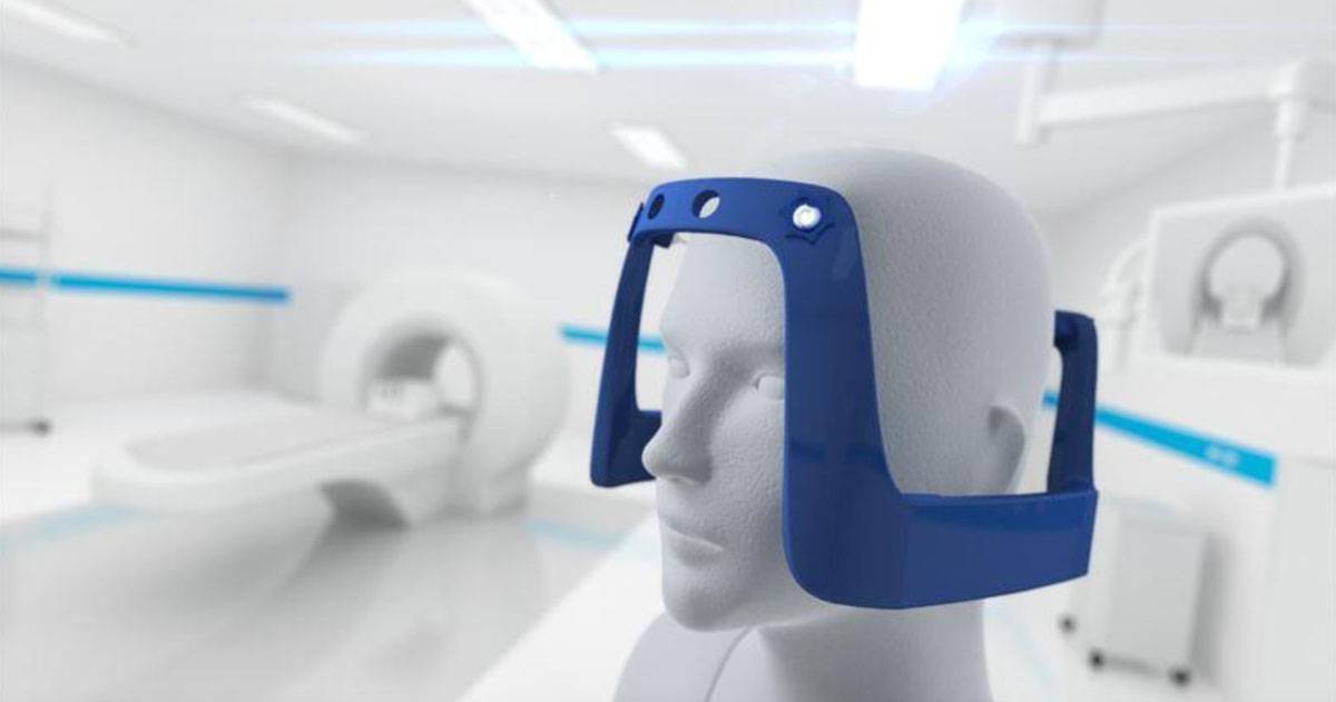
You may be offered stereotactic radiotherapy (SRT), which involves applying a strong dose of radiation from an external machine. The radiation beam is precisely targeted, to destroy the abnormal tissues and cause as little damage as possible to the surrounding tissue. One type of SRT is known as Gamma Knife surgery.

This does not involve a traditional surgical procedure with a knife. Rather, it is a method of applying a strong and highly focused dose of radiation from multiple angles at once. This maximises the deliberate damage done to the cancerous tissue, and minimises the risk of side effects and complications.

SCT is regarded as a safe and effective alternative treatment to open brain surgery. The patients may also be offered treatment with steroids or chemotherapy as a part of their treatment. 


The 2022 International Brain Tumour Awareness Week (IBTA) is taking place from 29 October to the 5 November. The aim is to raise awareness of brain tumours, and to help support anyone who has been impacted by a brain tumour, whether through a diagnosis for themself or a family member or friend.

The IBTA is organised by the International Brain Tumour Alliance, who are keen to increase the levels of research into brain tumours. Kathy Oliver, Founding Co-Director and Chair at IBTA, told the Brain Tumour Research charity: “This week is for everyone who has been impacted by a brain tumour.”

She added: “It is for anyone who hopes to see better outcomes for those diagnosed with this awful disease and – on a personal front – it is for Colin, my son who was diagnosed with a brain tumour in 2004 at age 24 and passed away, aged 32, in August 2011. I hope you are able to support our week.”

Suggested activities to help support IBTA include sharing stories about living with a brain tumour to local media, or with local community groups and schools. Other ideas include starting a blog about living with a brain tumour or caring for someone with a diagnosis, or developing educational resources such as webinars or online learning materials.

Popular fund-raising activities to help raise money for further brain tumour research include sponsored walks, fun runs, or quiz nights. The IBTA has also produced logos for supporters to download, and share on social media channels such as Facebook, Twitter, TikTok, and Instagram. 

IBTA have also organised a virtual exhibition titled Brilliance! which is designed to celebrate the creativity of the global brain tumour community, and is available to view on their website.


If you would like some information about gamma knife radiosurgery in the UK, please get in touch today. 


Gamma Knife surgery is a rather misleading term, because it isn’t surgery in the traditional sense, and doesn’t involve a knife. It involves precisely targeted narrow beams of low-dose radiation, which combine at the site of the tumour to destroy the cells. The exact position of the tumour is detected using scans to build accurate 3D images. 

This type of surgery is also known as stereotactic radiotherapy, or SRT. It has the advantage over conventional open surgery of substantially reducing the risk of bleeding and infection, and it has faster recovery times.

Gamma Knife surgery is less intrusive than open surgery, and the ability to precisely target the radiation beams means that the surrounding healthy tissue and organs will suffer little or no damage. It also means that you will usually need fewer treatments than with other types of radiotherapy, because a higher dose of radiation per treatment is used. 

This treatment is most suited to small well-defined tumours. The decision whether or not it is most suitable will be made on an individual basis by a medical professional, or a multi-disciplinary team. If the tumour is near important nerves which may be damaged by radiation, other types of surgery may be recommended. 

The most frequent type of tumours which are treated with gamma knife surgery are benign primary tumours, such as acoustic neuromas, pituitary adenomas and chordoma or meningiomas. Larger tumours are unsuitable for the treatment because the low doses of radiation involved wouldn’t be enough to destroy the tumour cells. 

Increasing the dose of radiation would present too higher risk of damaging the healthy brain tissue, so for larger tumours, or those with ill defined edges, Gamma Knife surgery would not be recommended. If there are multiple tumours present, then the treatment would also usually be unsuitable.


Researchers at the University of Bristol are developing a new blood test that could detect brain tumours at an earlier stage, the BBC reports. The test could be carried out at a GP surgery, avoiding the need for a patient to be transferred to a waiting list to see a consultant. 

The earlier a brain tumour is detected, the more effective and specific the treatment plan can be. The test will use mathematical models to examine and compare biomarkers for the most common types of brain tumour found in adults. 

Dr Johanna Blee said: “We are hopeful this research will ultimately aid the development of a simple blood test for brain tumours. Our findings provide the basis for further clinical data on the impact of lowering the current detection threshold, to allow earlier detection of GBMs using blood tests.” 

She added: “We have also demonstrated how our models can be combined with other diagnostics such as scans to enhance clinical insight with a view to developing more personalised and effective treatments.” 

The earlier a brain tumour is diagnosed, the more chance the patient has of being successfully treated. Biomarkers, or biological markers, are indicators such as a gene or molecule in the body that can be measured by clinicians to provide useful data.

For example, they can help determine what kind of tumour it is, and give a prediction about how fast it will grow. Medics may also use the test to determine which kind of treatment will be most effective. 

Research into biomarkers is still in the relatively early stages, and currently not all hospitals offer the tests. They do not always provide 100% reliable results, but they are useful for making a prognosis, and to help medics tailor a treatment plan that is most appropriate for the patient.

If you would like some information about gamma knife radiosurgery, please get in touch today.

There are different types of brain tumour, which are classed as malignant (cancerous) or benign (non-cancerous). They are further labelled according to the position in the brain, and graded from 1-4. Grade 1 and 2 are the least serious non-cancerous tumours, and grades 3-4 are cancerous. Here is some more information about how they are graded. 

Why are tumours graded?

Brain tumours are graded to provide you with an accurate diagnosis of your condition, and also to guide clinicians on a treatment pathway, and understand how the tumour might develop. The grading will usually be decided after a biopsy has been carried out, where a neuropathologist analyses the cell patterns from a sample of the tumour.

The more abnormal the brain tumour cells appear under a microscope, the higher the grade of tumour. Grade 1 tumours can have almost normal looking cells, while a grade 4 tumour will have the most abnormal looking cells. 

Sometimes, a tumour can be ‘mixed grade’ which means that the cells contained both low grade and high grade patterns. Even if the majority of the cells were low grade, the presence of high grade cells means that the tumour will be classed as high grade. 

Low grade tumours

A tumour that is grade 1 or 2 is classed as a low grade tumour. They are slow growing, and unlikely to spread or return if they are removed. Although they are benign, this doesn’t they are harmless or won’t require treatment. Depending on the position of the tumour in the brain, it can cause serious symptoms that may even be life threatening. 

Some benign tumours can cause a build up of pressure on the brain, by blocking the flow of cerebrospinal fluid which is essential for the brain to function normally. The tumour can also press on other areas of the brain, which may eventually cause damage. 

Furthermore, sometimes a low grade tumour can progress into a malignant grade 3 tumour, which could then develop into a grade 4 tumour. A grade 1 or 2 tumour may require surgery to remove it. 

High grade tumours

High grade tumours are graded as 3 or 4. They are fast growing and malignant (cancerous), and likely to spread to other areas of the brain or spinal cord. A combined treatment of surgery and radiotherapy or chemotherapy is usually required. 

Receiving your diagnosis

When your doctor or healthcare professional explains the grading of your brain tumour, don’t be afraid to ask questions, and ask them to explain anything that you don’t understand. They should be able to talk through all the recommended treatment options, for example.

Sometimes, certain hospitals or treatment centres run clinical trials of potentially effective new therapies. If this is something that you might be interested in taking part in, ask your doctor if there are any suitable programmes that you could sign up for. In some cases, you may also want to request a second opinion to confirm the diagnosis. 

If you would like some information about gamma knife radiosurgery, please talk to us today.

If you’ve been diagnosed with a brain tumour, or referred to a specialist by your GP for further testing, then no doubt you will have of lot of questions and new experiences and emotions to deal with. One of the things which often causes anxiety for new patients is going for a scan, but unfortunately multiple scans will usually be necessary. 

Here is some more information about the type of scans you will have, and some tips on how to feel calm and prepared during your scan. 

CT Scans

CT scans, or CAT scans as they are often called, refer to Computerised Tomography scans. The CT scanner will take multiple images of the inside of your head, and then computer technology will be used to stack them together into one 3D image. This allows clinicians to confirm if you have a brain tumour, and if so, where it is positioned and how big it is.

MRI scans

MRI stands for Magnetic Resonance Imaging, and this type of scanner uses magnetic fields rather than x-rays. However, the 3D image is created in a similar way, by taking several 2D images of the inside of your head, which are then used to build a single 3D image.

PET scans

PET scan stands for positron emission tomography scan. They work by injecting a slightly radioactive substance into the body, and a specialised camera is then used to highlight may areas of abnormality. They can provide a more accurate diagnosis of a tumour than MRI scans, but are more often used for monitoring purposes.

Before your scan

If you have told that you have an appointment for a scan, find out in advance which type of scan it will be, and ask your doctor to talk through the process with you. If you would find it helpful, record the conversation so you can play it back later and make sure that you have understood all the points. 

The scanning process

Both MRI and CT scans involve placing your head and neck inside the scanner. An MRI scanner is usually a tube shape, while a CT scanner is ring shaped. CT scans are usually fairly quick, taking between one to 10 minutes, depending on the model of scanner.

MRI scans usually take longer, and the time taken varies depending on how large the area being scanned is, and how much detail is required. It could be anything from 15 to 90 minutes. 

Before your scan, a clinician will conduct a health questionnaire to make sure that the procedure is safe for you. You’ll be asked to remove any metal items that you are wearing, such as jewellery, including piercings and watches. You will also need to remove dentures, hearing aids, glasses, and wigs. 

One of the main reasons that people fear scans is because they find the prospect claustrophobic. This is perfectly understandable and very common. It may help to have someone with you, such as a friend or relative. 

Let your GP or hospital staff know in advance if you think that you will have difficulty in coping because of your anxiety. They may prepare a mild sedative to help you feel calmer, although you should have someone to drive you home afterwards if this is the case. You will also be given an emergency button to press if you want to stop the scan at any point. 

In some cases, staff may be able to position mirrors which help you to see outside of the scanner, which can reduce feelings of claustrophobia. It may be useful to explore some mindfulness techniques, which focus on controlled breathing as a way of managing anxiety. 

Scans are not painful procedures, but they can be disconcerting experiences. An MRI scanner will make intermittent loud tapping noises, for example, which is created by the electric current being turned on and off. You’ll usually be given the option of listening to music through headphones, or given earplugs, which can help to keep you calm.

Remember that it’s important not to talk or move during the scan, as this will result in blurred and inaccurate images. Unless you have had a sedative, you should not experience any after effects from the scan. 

It usually takes one to two weeks for the results of your scan to be available. This can be an emotionally demanding time, and it may help to join some support groups which put you in touch will others in a similar situation. 

If you would like some more information about gamma knife surgery in the UK, please get in touch with us today. 

There has been a 30% rise in brain tumour diagnosis in Scotland over the last 20 years, the Daily Record reports. The latest figures from the health body show that there were 1,069 cases diagnosed in 2017-19, compared to 822 in 2000-2002.

Dr David Jenkinson, the Brain Tumour Charity’s chief scientific officer, said: “These worrying figures show just how urgently we need to act on this devastating and life-changing disease.”

He continued: “While brain tumours remain relatively rare, incidence has continued to rise significantly over the last two decades, and this has unfortunately not yet been matched by the tangible progress in diagnosis, treatment and survival outcomes seen in many other cancers.”

The Brain Tumour Charity campaigns to raise awareness of the disease, as well as supporting and funding new treatment centres. The earlier the condition is diagnosed, the better the chance of the patient receiving the most appropriate treatment plan, that could save or prolong their life.

The major symptoms include unexplained fits or seizures. This may involve uncontrollable jerking of the limbs, a spell of confusion or emotional disturbance, or complete loss of consciousness. Anyone experiencing these symptoms should go straight to A&E, where they may be referred for a brain scan.

Other common symptoms include frequent headaches which get progressively worse over time. However, headaches alone are not usually a sign of a brain tumour, unless they are accompanied by sickness and drowsiness, especially in the morning.

In some cases, a brain tumour may cause vision disturbances, such as lateral blind spots or flashing lights. It may also lead the sufferer to become more withdrawn and confused, and struggle with language and memory skills, which impedes on their day to day life.

If you would like some information about gamma knife surgery in the UK, please get in touch today.
The All-Party Parliamentary Group on Cancer (APPGC) has discussed the diagnosis and treatment of brain tumours at a recent meeting. The Brain Tumour Charity reports that one of their Involvement Champions attended the meeting at Westminster to talk about her personal experience.

Phillipa Anders recently lost her husband Rob, after he was diagnosed with a grade 4 brain tumour in 2020. She talked about the lack of personal support Rob received at the point of diagnosis, and advocated the use of Holistic Needs Assessments to make sure patients are given sufficient advice from their healthcare team.

Philippa also raised the wider point that brain tumours are often diagnosed at the point of emergency admission to hospital, unlike many other types of cancer which are now detected by doctors much earlier. As brain tumours are responsible for the most cancer deaths in people under 40, and children, earlier diagnosis could save many lives.

Symptoms of a brain tumour include headaches, which are often combined with a frequent feeling of sickness, and problems with vision, such as the appearance of flashing lights, blurred vision, tunnel vision, or blind spots. Around eight out of 10 people will experience seizures, and some people may feel drowsy or lose consciousness.

Philippa told the meeting: ‘It is essential that brain tumours are diagnosed at the earliest possible opportunity to allow referrals and support to be put in place, but brain tumours aren’t ‘staged’ in the same way as other cancers and this needs to be factored into the NHS targets and ambition.”

She added: “The 10-Year Cancer Plan should include greater awareness and understanding of signs and symptoms, in healthcare settings, work settings, and the broader community.”

The APPGC also discussed the need for better communication between patients and healthcare professionals, better IT and digital systems, and improved staff retention rates within the NHS.

If you would like some information about gamma knife surgery in the UK. please get in touch today.

Research into the use of Gamma Knife to treat primary brain tumours is ever evolving. In August 2022 the Journal of Neurosurgery (JNS) published the results of a feasibility study in a clinical article called, ‘Conventionally fully fractionated Gamma Knife Icon re-irradiation of primary recurrent intracranial tumors: the first report indicating feasibility and safety’.

Ian Paddick, Chief Physicist for Amethyst UK comments on the recently published article. He said; “This collection of case studies demonstrates the feasibility and potential safety of using Gamma Knife to deliver conventionally fractionated salvage treatments to recurrent tumours close to organs at risk that have received doses close to, or up to full tolerance.

“In addition, the article goes on to remind us that we have animal and retrospective clinical data that shows that brain tissue can recover significantly after irradiation – perhaps 75% in two years. This allows for reirradiation. Therefore, there may be room to be more aggressive when clinically needed.”

Why use Gamma Knife for this sort of treatment?

Gamma Knife is ideally suited to irradiation of complex targets to low doses. It can, for example, deliver complex treatment plans in 2Gy fractions in around 10 minutes, making it as efficient as a Linac. The feasibility study highlighted in the JNS article used a median number of 28 fractions. Ian Paddick concluded; “Gamma Knife’s excellent conformity and gradient is better suited to target tumours surrounded by previously irradiated tissue.”

Cerebral arteriovenous malformations (AVMs) arise when an abnormal tangle of blood vessels occurs in the brain as a result of an abnormal connection between an artery and vein. These can have serious consequences such as haemorrhaging, stroke, brain damage and seizures. Gamma Knife radiosurgery represents a massive improvement in the treatment options for AVMs, as a targeted and precise, non-invasive approach.

What are the treatment options available for AVMs?

The three treatment options are endovascular embolization (“gluing”), neurosurgical resection (surgery) and stereotactic radiosurgery (Gamma Knife). In some cases, just one treatment modality is required but a mix of these treatments can sometimes also be used depending on the characteristics of the AVM and the patients’ own preferences.

Generally, for smaller AVMs that are under 3cm in size, Gamma Knife treatment is commonly used. Embolization can be used to de-vascularise or shrink the AVM prior to surgical or radio-surgical treatments.

How are AVMs diagnosed?

AVMs can occur anywhere in the body but most commonly present in the brain). Many patients have no symptoms of an AVM until a bleeding event occurs. AVMs are usually diagnosed by MRI or CT head. Angiography can be performed to give the best and most detailed picture of the vascular anatomy. They can often be discovered incidentally, that is to say when a scan is done for another reason. Alternatively, AVMs can present with symptoms such as seizures, headaches or a neurological problem like weakness in one part of the body or speech disturbance. AVMs can therefore have a significant impact on patient’s lives and the specifics of each case should be discussed with a skilled and experienced team.

Why do we treat arteriovenous malformations (AVMs)?

Untreated, AVMs carry a risk of haemorrhaging which can cause brain damage or death. A 2020 study on the clinical outcomes following cerebral AVM haemorrhage published by the National Library of Medicine found that an AVM rupture has around 20% likelihood to result in mortality, 45% likelihood to result in a minor or major deficit, and 35% likelihood of complete recovery. This means that even when an AVM presents without symptoms and a bleeding event has not occurred, it can be important to treat them.

There are also several factors that can increase the risk of AVMs haemorrhaging, including its size. Although it seems counterintuitive, smaller lesions tend to carry a higher risk of haemorrhage due to the higher arterial pressure that builds in smaller vessels.

What are the benefits of Gamma Knife?

Gamma Knife stands out as a treatment option from surgery and embolization in a number of ways. It mitigates the many risks associated with patients going under general anaesthetic, and of open surgery such as infection, bleeding, or wound problems.

Gamma Knife also has the capability to treat very deep and sensitively located AVMs which cannot be otherwise treated. This is because it uses up to 192 precisely focussed beams of radiation to target selected brain lesions, without harming the surrounding healthy brain tissue. From this, over time the walls of the AVM thicken and scar, eventually closing most of the vessels supplying the AVM, thus preventing rupture. Although the procedure involves several steps on the day of treatment including imaging and planning, the treatment itself in many cases can take less than an hour.

Article by Ms. Mary Murphy Clinical Director of Neurosurgery at The National Hospital for Neurology and Neurosurgery. Quality and safety lead for the specialist hospital board at University College Hospital London.